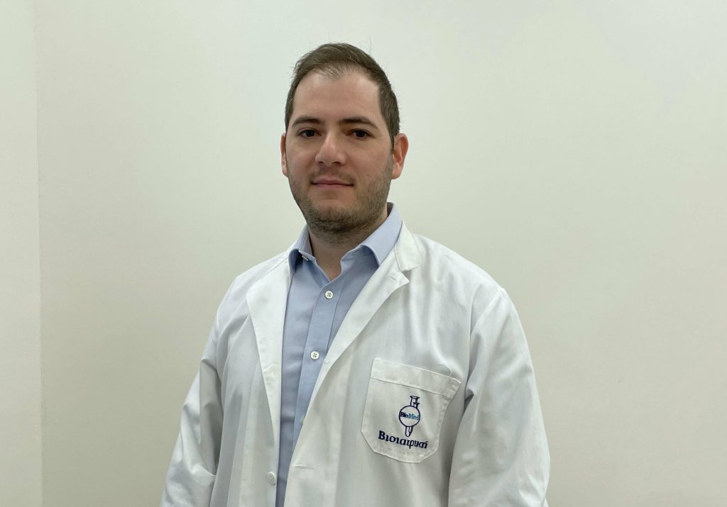
Dr Andreas Panagiotou* answers the most frequently asked questions about the peculiarities of joint imaging with MRI.
What is MRI Arthrography?
MRI arthrography is one of the internationally recognized imaging methods for the evaluation of any kind of joint pathology. In other words, it is an imaging test that uses a contrast agent to better evaluate the joint (e.g., the shoulder, knee or hip), which cannot be achieved through standard MRI alone.
In MRI arthrography, a fine needle is used to inject a contrast agent into the joint, followed by an MRI scan to obtain better and more accurate images of the joint. While MRI arthrography is most commonly used to examine the shoulder and hip joints, it can also be used to examine other joints such as the knee, wrist, ankle or elbow.
Why I may need MRI arthrography?
The test can be performed on a joint either when there is persistent and unexplained pain, discomfort, loss of motion and/or changes in the way the joint functions, or when the issue is not properly addresses with a standard MRI imaging.
The most common reasons are:
– The evaluation of pathological issues (e.g., tears/ruptures) in the soft tissues of the joint, such as cartilage, ligaments, tendons, cartilage, and joint capsules.
– Checking for damage from repeated dislocations of the joint.
How is MRI Arthrography performed?
MRI arthrography is an invasive medical procedure, so it requires a high degree of expertise from the medical radiologist. In direct arthrography, which offers the best possible imaging result, the joint is catheterized with a fine needle, guided by ultrasound (US) or computed tomography (CT), in order to inject the contrast agent directly into the joint cavity. After the contrast agent is injected into the joint, an MRI scan follows.
Although this is a specialized test, no preparation is required from the patient prior to the test. During and shortly after the injection there may be some discomfort in the joint, however, it is temporary, and the subject can usually return to normal activities immediately after the procedure.
*Specialist Radiologist
Alpha Evresis Diagnostic Center, Bioiatriki Healthcare Group in Cyprus


